Saturday, April 10, 2010
Aneurysm presentation-- random clinical pearls
Diagnosis
1. Of patients with the worst headache of their life, ten percent have aneurysms
2. Sensitivity of LP to detect aneurysms decreases by seven percent per day
3. CT-A removes need for catheter angiography in those with more than 5 rbc's
4. SAH- aneurysmal peaks in April and September and nadirs in June and July
5. Population prevalence of aneurysm in 2 %
Treatment
6. **FENESTRATION OF LAMINA TERMINALIS IS EASY, DECREASES HYDROCEPHALUS INCIDENCE FROM 13 TO 2 PERCENT AND ALLOWS LUMBAR DRAINS
7. EEG is a "pseudoexam " under anesthesia
8. St Julien NSURG 2008 cardiopulmonary bypass without a chest incision (endovascular)allows fine control of BP and avoids circulatory arrest, hypothermia improves outcomes ( outcome of St Julien) Grade 0 , 1 (1.5 %), 2 (6.2%), 3 (12.1%), 4 (17.4 %). CP bypass is good for giant aneurysms
9. Fisher scale stratifies the risk of vasospasm . Grade 1: no blood Grade 2: vertical layer < 1mm
Grade 3: vertical layer > 1 mm, local clot Grade 4: ICH/IVH but minimal or no SAH. GRADE THREE IS MAXIMAL RISK> GRADE FOUR. Risk of vasospasm is 23 %. With modified Fisher scale, vasospasm is greatest with grade 4. About 20-30 % of vasospasms stroke.
10. Risk of rebleed is 4 % in first 24 hours, then 1-2 % per day for 4 weeks. Cumulative risk is 20 % at 2 weeks, 30 % at one month, 40 % at 6 months. Ventriculostomy which otherwise can be lifesaving also can precipitate a rebleed.
10. Risk of rebleed is


Hemorrhagic shock plus TBI no longer uniformly fatal
Combination is less lethal than formerly provided that CPP is maintained. Treatment algorithm: stop bleeding (factor 7), restore volume (whole blood, crystalloid), saline, plasma, platelets, pressors (phenylephrine, vasopressin, norepinephrine, DA), prophylactic phenytoin, for 7 days, treat fevers with tylenol, aggressive nutrition, use hemicraniectomy for impending herniation is efficacious, use GCS to communicate.
Prehospital care: avoid hypoxia-- give oxygen, avoid hypotension, hypertonic saline is good; mannitol only if intravascular volume can be maintained, generally avoid hyperventilation unless herniating.
ICP monitor if GCS < 8 and abnormal CT scan. Want ICP< 20, intervention threshold around 25. ICP plateau or A waves are sine qua non of herniation. These are best seen with changing sweep speed on monitor to minutes. B waves last from 0.5 to 2 minutes and are associated with changing brain compliance not increased ICP. C waves are ICP waves associated with respiration.
Components of a "brain code" are 1) elevated HOB to 45 degrees 2) HV to pCO2 around 35 3) mannitol .5 grams/kg 4) saline bullet (see http://neurologyminutiae.blogspot.com/2010/04/saline-bullets-and-hypertonic-saline.html) 5) CSF drainage
from lecture by Dr Ling
Vasospasm after traumatic brain injury
Unlike its better known couin that occurs after SAH, vasospasm after TBI follows a different time course of 10-21 days is often subclinical and is best treated with nicardipine and endovascular therapy. It can be monitored with TCD.
Source-- lecture Col. Geoffrey Ling , MD
Saturday, April 03, 2010
Pearls on blood pressure, misc and hemorrhagic stroke care
1. PET studies do not support the concept of an ischemic penumbra, hence blood pressure control should be used judiciously (Schellinger et al, Stroke 2003)
2. The occurrence of ICH is strongly related to prevailing blood pressure, however no definitive evidence exists that recurrent ICH in the acute setting relates to blood pressure or control thereof (Jauch et al. Stroke 2006)
3. Intracranial hypertension is associated with a worse outcome
4. Prior statin use is associated with decreased perihemorrhage edema and decreased 30 day mortality ; however this data is retrospective (Naval et al., 2 refs Neurocritical Care 2008)
5. The Stroke Council continues to advocate for 2-4 weeks of prophylactic antiepileptic therapy in patients with SICH and SAH
6. Hematoma size (Stoke 1997, Brott et al) and growth (Davis et al, Neurology 2006) are correlated with mortality
7. The "spot sign" or contrast extravasation in CTA may identify patients at high risk of hematoma expansion
8. ICH < 30 cc may benefit from intraclot alteplase
9. MIS minimal invasive surgery is also considered under investigation although certain types of ICH do not benefit
Sunday, March 28, 2010
Carotid artery webbing
case report at AAN 2010 also known as "atypical FMD" a 41 year old woman with headache for months, facial droop and aphasia cutely. MRI showed an M1 occlusion, and delayed time to peak in entire MCA. Catheter showed bilateral ICA webbing with stagnant flow. No standard of care is known.
MCA arrow sign in MCA aneursmal SAH
In a review, arrow sign was present in 4 patients with SAH all Fisher 3. That is 16 % of total MCA aneurysms and was not seen in any other type of aneurysm.
below is an arrow sign for an MCA trifurcation aneurysm (from neurology.org)


below is an arrow sign for an MCA trifurcation aneurysm (from neurology.org)


Ptosis and astasia with thalamic infarcts: case report (s)
Jain D. et al. Cerebral ptosis and astasia "Lateral pulsion" due to a left anterior thalamic lesion. Mechanisms are reviewed.AAN 2010:PO2:102
Alderazi Y. Thalamic infarction causing astasia-abasia, ataxia and asterixis. clinical and radiological features of two cases. PO2:108. Wide based gait past pointing and intention tremor on right, with left posterolateral thalamic infarct. Second case with left arm drift, left asterixis, inability to stand unassisted with right lateral thalamic acute stroke and old left cerebellar hemorrhage.
Alderazi Y. Thalamic infarction causing astasia-abasia, ataxia and asterixis. clinical and radiological features of two cases. PO2:108. Wide based gait past pointing and intention tremor on right, with left posterolateral thalamic infarct. Second case with left arm drift, left asterixis, inability to stand unassisted with right lateral thalamic acute stroke and old left cerebellar hemorrhage.
Sneddon's syndrome need for angiography
Faris et al. Sneddon's syndrome without antiphospholipid antibodies: a report of 26 cases with cerebral angiography (Rabat). Neurology 2010 74:9:PO2:099.
Authors emphasize Sneddon's syndrome (stroke plus livedo racemosa) is NOT identical to APL syndrome. 26 patients were studied retrospectively with a combination of focal motor deficits and dementia. Imaging always showed infarcts with white matter involvement. Angio showed a distal arteriopathy in 18 cases with pial networks in cases. Two had hematomas. The authors suggested angiography to prevent the unwarranted and dangerous potential use of anticoagulation.
Authors emphasize Sneddon's syndrome (stroke plus livedo racemosa) is NOT identical to APL syndrome. 26 patients were studied retrospectively with a combination of focal motor deficits and dementia. Imaging always showed infarcts with white matter involvement. Angio showed a distal arteriopathy in 18 cases with pial networks in cases. Two had hematomas. The authors suggested angiography to prevent the unwarranted and dangerous potential use of anticoagulation.
Friday, March 26, 2010
NSE after cardiac arrest during hypothermia predicts outcome
AN 2010 Po1.048 JEF Fugate, Wijdicks et al. NSE was measured serially in comatose patients undergoing hypothermia. A cutoff was used of 33 ug/L. NSE was measured at day one and day three, with higher NSE suggesting poor prognosis (defined as one year mortality). 48 patients, 41 had first day NSE, 14 had third day NSE. For first day NSE, the number with high level is given with number of survivors at one year in parenthesis 19 (3) with a low NSE being 22 (11). Trend in NSE on day 3 was highly predictive (p<.015). The sensitivity for first day NSE was 59 % for one year mortality with improvement to 66 % if 3 day NSE is included.
Thursday, March 25, 2010
Four score is predictor of outcome in coma after cardiac arrest
Neurology AAN 2010 PO1.045 Wijdicks et al.
The Four Score differs from the GCS because it has 4 components-- eye, motor, brainstem and respiration (latter two are not included in GCS) . Prospectively looked at patients from 2006-2009 (n=131) and looked at outcome after one year. 91 died. 31 had four score less than 4 at day one and of these, zero survived. Of patients with GCS of 3, 4 (7 %) survived at one year. The Four Score had a specificity for absent survival at one year of 100 % versus 90 % for GCS of 3.
Description of the FOUR Score
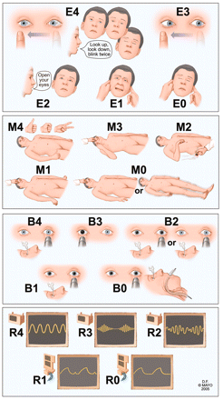
The FOUR score has 4 components: eye responses, motor responses, brainstem reflexes, and respiration pattern. Each component has a maximal value of 4 (Figure 1). Assessing all components of this score usually takes only a few minutes.5 The eye response component of the FOUR score allows differentiation between a vegetative state (eyes open but do not track) and a locked-in syndrome (eyes open, blink, and track vertically on command). The motor assessment component of the FOUR score combines the withdrawal reflex and decorticate rigidity responses because these conditions are often difficult to distinguish clinically. The motor component includes a complex command (the patient is asked to produce a thumbs-up hand signal, a fist, and the peace sign) that determines whether patients are alert.7 Similarly, the motor component of the FOUR score can detect signs of severe cerebral dysfunction, such as myoclonic status epilepticus. Such dysfunction is often a poor prognostic sign for patients with suspected anoxic brain injury.8 The brainstem components of the FOUR score assess the pons, the mesencephalon, and the medulla oblongata in various combinations. The FOUR score also includes an assessment of Cheyne-Stokes respiration and irregular breathing; such signs can indicate bihemispheric or lower brainstem dysfunction of respiratory control. For patients who have undergone intubation, the FOUR score records the presence or absence of a respiratory drive.
FIGURE 1.
Description of Full Outline of UnResponsivenes (FOUR) score. Eye response: E4 = eyelids open or opened, tracking, or blinking to command; E3 = eyelids open but not tracking; E2 = eyelids closed but open to loud voice; E1 = eyelids closed but open to pain; E0 = eyelids remain closed with pain. Motor response: M4 = thumbs-up, fist, or peace sign; M3 = localizing to pain; M2 = flexion response to pain; M1 = extension response to pain; M0 = no response to pain or generalized myoclonus status. Brainstem reflexes: B4 = pupil and corneal reflexes present; B3 = one pupil wide and fixed; B2 = pupil or corneal reflexes absent; B1 = pupil and corneal reflexes absent; B0 = absent pupil, corneal, and cough reflex. Respiration pattern: R4 = not intubated, regular breathing pattern; R3 = not intubated, Cheyne-Stokes breathing pattern; R2 = not intubated, irregular breathing; R1 = breathes above ventilatory rate; R0 = breathes at ventilator rate or apnea.
Sunday, March 21, 2010
The Broken Heart Syndrome
llan Wittstein has published and given lectures and states there are clinical criteria, diagnostic criteria and treatment and prognosis information that can be readily identified. Synonyms include neurogenic stunned myocardium, acute coronary syndrome, stress myocarditis, and Takotsubo syndrome (named after the japanese pot used to capture octopus). The syndrome is a REVERSIBLE disorder with very abnormal EKG, U waves, ST elevations, Q waves, elevated troponins, normal coronaries on cath, and return of EF to normal within days to weeks. The pathology includes contraction band necrosis in myocardium, and is linked to hypersympathetic state. It is a huge problem involving 2 percent of patients undergoing cath and 5-7 % of women. Most patients are postmenopausal Caucasian women with risk factors who present with chest pain and shortness of breath. Many have mood disorders
Diagnostic criteria are divided into "helpful" and "required"
HELPFUL-
1. Acute trigger-- could be emotional (anxiety, joy, grief, fear, anger) or physical (procedure, respiratory drugs) even surprise party.
2. Characteristic EKG-- presenting EKG has steep ST elevation without reciprocal changes, T wave inversions everywhere, QT prolongation, that becomes milder within 2-4 days
3. Troponin elevation is mild-- less than 5, never more than 20
REQUIRED
1. Absent coronary thrombosis
2. Wall motion abnormalities extend beyond a single coronary artery territory ( 3 patterns: apical, basal, and midventricular)
3. Rapid recovery of systolic function within 2 weeks at most
Diagnostic tests that are helpful (but possibly hard/unlikely to obtain esp. acutely)
1. MRI heart unlike ECHO differentiates dead and stunned tissue. Dead cardiac tissue lights up with Gadolinium but stunned heart will not
Therapy:
1. supportive-- possibly not in ICU- arbs, ACEi's, diuretics. Anticoag if apex not moving to prevent clot kicking, avoid pressors (catechols are a problem)
2. Balloon pump better than pressors
3. HHH good for brain, bad for heart
Prognosis
1. recurrence 3-10 percent with 2 % mortality
2. Death is due to etiology not to cardiac dysfunction per se.
Pathophysiology
contraction band necrosis- direct myocyte injury related to calcium overload.
Diagnostic criteria are divided into "helpful" and "required"
HELPFUL-
1. Acute trigger-- could be emotional (anxiety, joy, grief, fear, anger) or physical (procedure, respiratory drugs) even surprise party.
2. Characteristic EKG-- presenting EKG has steep ST elevation without reciprocal changes, T wave inversions everywhere, QT prolongation, that becomes milder within 2-4 days
3. Troponin elevation is mild-- less than 5, never more than 20
REQUIRED
1. Absent coronary thrombosis
2. Wall motion abnormalities extend beyond a single coronary artery territory ( 3 patterns: apical, basal, and midventricular)
3. Rapid recovery of systolic function within 2 weeks at most
Diagnostic tests that are helpful (but possibly hard/unlikely to obtain esp. acutely)
1. MRI heart unlike ECHO differentiates dead and stunned tissue. Dead cardiac tissue lights up with Gadolinium but stunned heart will not
Therapy:
1. supportive-- possibly not in ICU- arbs, ACEi's, diuretics. Anticoag if apex not moving to prevent clot kicking, avoid pressors (catechols are a problem)
2. Balloon pump better than pressors
3. HHH good for brain, bad for heart
Prognosis
1. recurrence 3-10 percent with 2 % mortality
2. Death is due to etiology not to cardiac dysfunction per se.
Pathophysiology
contraction band necrosis- direct myocyte injury related to calcium overload.
air embolism and air travel
The Neurologist 2010 A 62 year old woman with cerebral artery air embolism during commercial air travel. 16: 136-137.
Notes
usual list of associations with air embolus-- surgery, scuba diving, induced abortion, angiography and pneumothorax, orogenital sex on a woman with cerebral air emboli (see Crit Care Med 1988).
A new one is air travel. In this case, pulmonary bullae due to emphysema occurred as the bullae expanded as the pressure in the cabin was reduced, leading to rupture of the bullae, pneumothorax and air embolism. Barotrauma was presumably the proximate cause.
Pearls on pediatric strokes
hat tip to Lori Jordan MD JHU
1. Strokes in kids are as common as brain tumors, about 2-3/100,000
2. In children ICH = bland infarcts, different than adults (Fullerton, Neurology, 2003). ICH is often due to AVM's
3. Among bland infarcts, 25-35 % are cardioembolic, 25 % are dissections, other unusual causes include moya moya, sickle cell disease, HIV and varicella (not in order).
4. Subarachnoid hemorrhage in kids is usually aneurysmal
5. Kids at risk often have an inciting event such as trauma or surgery
6. Kids have a high risk of delay in diagnosis
7. Sicklers have 10 % stroke, but 20 % more of silent stroke; with SCA and CVA stat consult Hematology for transfusion
8. Many barriers to alteplase use exist, including diagnosis, , lack of evidence and mimics, and delays, but document why alteplase is not given
9. MERCI and multi MERCI are not studied in kids
10.AHA guidelines for pediatric stroke published Stroke 2008
11. Presentation in children is much more likely to include seizure (25 % v. 5 % in adults)
12 . Suggested eval: MRI, MRA H/N, hypercoagulability workup complete, TTE/bubble, HB electropheresis, HIV,
13 references
Roach ES, Golomb MR, Adams R, Biller J, Daniels S, Deveber G, et al.
Management of stroke in infants and children: A scientific statement from a
special writing group of the american heart association stroke council and the
council on cardiovascular disease in the young. Stroke 2008;39:2644-91.
• Amlie-Lefond, C. et al. Use of alteplase in childhood arterial ischaemic stroke: a
multicentre, observational, cohort study. 2009: Lancet Neurol. 8, 530-536.
• Jordan LC, Johnston SC, Wu YW, Sidney SS, Fullerton HJ. The importance of
cerebral aneurysms in childhood hemorrhagic stroke: a population-based study.
Stroke 2009;40:400-405.
• Beslow LA, Licht DJ, Smith SE, Storm PB, Heuer GG, Zimmerman RA, Feiler
AM, Kasner SE, Ichord RN, Jordan LC. Predictors of outcome in childhood
intracerebral hemorrhage: a prospective consecutive cohort study. Stroke 2009;
1. Strokes in kids are as common as brain tumors, about 2-3/100,000
2. In children ICH = bland infarcts, different than adults (Fullerton, Neurology, 2003). ICH is often due to AVM's
3. Among bland infarcts, 25-35 % are cardioembolic, 25 % are dissections, other unusual causes include moya moya, sickle cell disease, HIV and varicella (not in order).
4. Subarachnoid hemorrhage in kids is usually aneurysmal
5. Kids at risk often have an inciting event such as trauma or surgery
6. Kids have a high risk of delay in diagnosis
7. Sicklers have 10 % stroke, but 20 % more of silent stroke; with SCA and CVA stat consult Hematology for transfusion
8. Many barriers to alteplase use exist, including diagnosis, , lack of evidence and mimics, and delays, but document why alteplase is not given
9. MERCI and multi MERCI are not studied in kids
10.AHA guidelines for pediatric stroke published Stroke 2008
11. Presentation in children is much more likely to include seizure (25 % v. 5 % in adults)
12 . Suggested eval: MRI, MRA H/N, hypercoagulability workup complete, TTE/bubble, HB electropheresis, HIV,
13 references
Roach ES, Golomb MR, Adams R, Biller J, Daniels S, Deveber G, et al.
Management of stroke in infants and children: A scientific statement from a
special writing group of the american heart association stroke council and the
council on cardiovascular disease in the young. Stroke 2008;39:2644-91.
• Amlie-Lefond, C. et al. Use of alteplase in childhood arterial ischaemic stroke: a
multicentre, observational, cohort study. 2009: Lancet Neurol. 8, 530-536.
• Jordan LC, Johnston SC, Wu YW, Sidney SS, Fullerton HJ. The importance of
cerebral aneurysms in childhood hemorrhagic stroke: a population-based study.
Stroke 2009;40:400-405.
• Beslow LA, Licht DJ, Smith SE, Storm PB, Heuer GG, Zimmerman RA, Feiler
AM, Kasner SE, Ichord RN, Jordan LC. Predictors of outcome in childhood
intracerebral hemorrhage: a prospective consecutive cohort study. Stroke 2009;
Wednesday, March 17, 2010
CAA with vasculitis and edema responsive to steroids references
inflammation related CAA as described in : Ann Neurol 2004; 55: 250-256; Brain 2005; 128: 500-515; Neurology 2007; 68 (17); 1411-1416.
Sunday, March 07, 2010
ISC Abstract highlights 2010 San Antonio (pruned and edited)
1. Restrepo et al. (UCLA) Stroke pretreatment screening for fast Mag trial, involved a 90 second screen with a neurologist, focused, 72 % of patients so diagnosed had acute ischemic stroke, 24 % ICH, rest other
2. Albright et al. (Penn) studied the potential for the use of air ambulances to increase availability of services and found The combination of pre-hospital regionalization & air ambulance transport of acute stroke
patients would reduce the 135.7 million Americans without 60 minute access to a PSC by
half, to 62.9 million.
3. Kleindorfer et al. (Cincinatti) stratified t-PA eligibility by age and found contrary to hypothesis, the eligibility for rt-PA significantly increased with increasing age.
Age-Based Eligibility for and Treatment with Rt-PA
Age of Pt # Patients % Eligible for rt-PA % of Eligible Treated
18–44 97 4 (4.1%) 2 (50.0%)
45–54 219 15 (6.8%) 5 (33.3%)
55–64 320 21 (6.6%) 12 (57.1%)
65–74 392 32 (8.2%) 20 (62.5%)
75–84 502 47 (9.4%) 23 (48.9%)
85 300 29 (9.7%) 10 (34.5%)
Total 1830 148 (8.1%) 72 (48.6%)
4. Riccio et al (Buenos Aires) Occult v. non -occult AF compared in TIA and AIS. Age, female gender and left atrial area (LAA) are traditional determinants of AF. Out of 194 patients, there were 36 with known AF and 24 with occult AF. Patients with occult AF were younger, showed a higher proportion of males, had
a smaller LAA, and had more severe strokes. Traditional determinants of AF were associated
with known AF.Diabetes was associated with occult AF.
5. Gupta et al. (multicenter) General anesthesia during stroke resulted in worse outcomes.
2. Albright et al. (Penn) studied the potential for the use of air ambulances to increase availability of services and found The combination of pre-hospital regionalization & air ambulance transport of acute stroke
patients would reduce the 135.7 million Americans without 60 minute access to a PSC by
half, to 62.9 million.
3. Kleindorfer et al. (Cincinatti) stratified t-PA eligibility by age and found contrary to hypothesis, the eligibility for rt-PA significantly increased with increasing age.
Age-Based Eligibility for and Treatment with Rt-PA
Age of Pt # Patients % Eligible for rt-PA % of Eligible Treated
18–44 97 4 (4.1%) 2 (50.0%)
45–54 219 15 (6.8%) 5 (33.3%)
55–64 320 21 (6.6%) 12 (57.1%)
65–74 392 32 (8.2%) 20 (62.5%)
75–84 502 47 (9.4%) 23 (48.9%)
85 300 29 (9.7%) 10 (34.5%)
Total 1830 148 (8.1%) 72 (48.6%)
4. Riccio et al (Buenos Aires) Occult v. non -occult AF compared in TIA and AIS. Age, female gender and left atrial area (LAA) are traditional determinants of AF. Out of 194 patients, there were 36 with known AF and 24 with occult AF. Patients with occult AF were younger, showed a higher proportion of males, had
a smaller LAA, and had more severe strokes. Traditional determinants of AF were associated
with known AF.Diabetes was associated with occult AF.
5. Gupta et al. (multicenter) General anesthesia during stroke resulted in worse outcomes.
Sunday, February 07, 2010
Infective endocarditis and stroke: pearls
. The classic triad of infective endocarditis and stroke of fever, murmur and acute neurologic deficit is uncommon, occurring in less than half of patients, with murmur occurring in less than one third (due to decline of valvular disease as a cause) ; fever, embolism and high sed rate may also be seen in NBTE, arteritis with PAN, other rheumatologic disease, or atrial myxoma.
2. Organisms are more diverse than previously with Streptococcus representing only 60 percent, including resistant Group D Strep viridans (enterococcus faecalis) and Strep bovis (associated with GI neoplasia). Staph aureus is seen in up to 30 % especially those with i-v drug abuse, recent surgery, and no preexisting valve lesion. Patients with prosthetic valves may get S aureus and S epidermidis. Others, including immunocompromised may get HACEK bacteria and fungi ( hemophilus, actinobacillus, cardiobacterium, Eikinella, Kinzella). Among pretreated groups, culture negative disease has increased to 5.5 %.
3. Early onset (at presentation of first 48 hours) of neurologic symptoms is more common with Staph aureus than with streptococcal infections, that can occur late (54 v 19 %). Late embolism is especially common among patients with prosthetic valves (14/15 late strokes in one series had prosthetic valves). In native valve endocarditis, anticoagulation is of no benefit , certainly for at least 48 hours or until infection is controlled.
4. Cardiac vegetations initially larger than 10 mm are high risk, and are best seen with TEE rather than TTE. Embolic rate is much higher in presence of visible lesions, and vice versa, visible lesions are much more common among patients with detectable emboli. The 10 mm size may "open the debate" about the need for valve replacement.
5. The discovery of endocarditis without emboli does not dictate the cessation of otherwise needed anticoagulant therapy.
6. If cardiac surgery is needed, timing is dictated by common sense. One such protocol is to wait at least five days (until the edema of the stroke has settled) before considering operation, if possible.
7. Use heparin fairly early on (certainly within 48 hours) of stroke with prosthetic valves with endocarditis, especially if subclinical INR was found on presentation. Discontinue anticoagulation if possible in most cases of fungal endocarditis.
8. Intracranial hemorrhage (3-6 % of patients) occurs with aneurysm rupture, septic arteritis, conversion of a bland infarct, and late effects of immune deposition. Septic bacterial aneurysms may occur at distal branch points, but mycotic aneurysms, large ones, may occur proximally. However, one study suggested the vast majority of patients with aneurysms had abnormal CT scan. Present blood on CT or (if headache is present) pleocytosis on CSF examination weighs towards four vessel angiogram.
9. Serial angiography is indicated for mycotic aneurysms which may heal with antibiotics.
10. Special concern exists among patients with line induced sepsis and bland infarction that could result in late S Aureus superinfection of an initially bland infarct. Full infective endocarditis treatment regimen may be warranted in these patients and TEE may alternatively show evidence of vegetations.
Unknown angles
1. Role of MRA/ CTA
2. When surgery is required
2. Organisms are more diverse than previously with Streptococcus representing only 60 percent, including resistant Group D Strep viridans (enterococcus faecalis) and Strep bovis (associated with GI neoplasia). Staph aureus is seen in up to 30 % especially those with i-v drug abuse, recent surgery, and no preexisting valve lesion. Patients with prosthetic valves may get S aureus and S epidermidis. Others, including immunocompromised may get HACEK bacteria and fungi ( hemophilus, actinobacillus, cardiobacterium, Eikinella, Kinzella). Among pretreated groups, culture negative disease has increased to 5.5 %.
3. Early onset (at presentation of first 48 hours) of neurologic symptoms is more common with Staph aureus than with streptococcal infections, that can occur late (54 v 19 %). Late embolism is especially common among patients with prosthetic valves (14/15 late strokes in one series had prosthetic valves). In native valve endocarditis, anticoagulation is of no benefit , certainly for at least 48 hours or until infection is controlled.
4. Cardiac vegetations initially larger than 10 mm are high risk, and are best seen with TEE rather than TTE. Embolic rate is much higher in presence of visible lesions, and vice versa, visible lesions are much more common among patients with detectable emboli. The 10 mm size may "open the debate" about the need for valve replacement.
5. The discovery of endocarditis without emboli does not dictate the cessation of otherwise needed anticoagulant therapy.
6. If cardiac surgery is needed, timing is dictated by common sense. One such protocol is to wait at least five days (until the edema of the stroke has settled) before considering operation, if possible.
7. Use heparin fairly early on (certainly within 48 hours) of stroke with prosthetic valves with endocarditis, especially if subclinical INR was found on presentation. Discontinue anticoagulation if possible in most cases of fungal endocarditis.
8. Intracranial hemorrhage (3-6 % of patients) occurs with aneurysm rupture, septic arteritis, conversion of a bland infarct, and late effects of immune deposition. Septic bacterial aneurysms may occur at distal branch points, but mycotic aneurysms, large ones, may occur proximally. However, one study suggested the vast majority of patients with aneurysms had abnormal CT scan. Present blood on CT or (if headache is present) pleocytosis on CSF examination weighs towards four vessel angiogram.
9. Serial angiography is indicated for mycotic aneurysms which may heal with antibiotics.
10. Special concern exists among patients with line induced sepsis and bland infarction that could result in late S Aureus superinfection of an initially bland infarct. Full infective endocarditis treatment regimen may be warranted in these patients and TEE may alternatively show evidence of vegetations.
Unknown angles
1. Role of MRA/ CTA
2. When surgery is required
Tuesday, February 02, 2010
Percentage of patients with stroke with prior TIA
Hackam DG, Kapral MK, Wang JT et al. Most stroke patients do not get a warning: a population based cohort study. Neurology 73: 1074-1075, 2008.
cites data suggesting 17 % of CVA patients have prior TIA. Authors review 16, 409 charts. Timing of TIA is not addressed. 12.4 % had prior TIA, but 20% of those with large arter TIA's, much lower with hemorrhagic stroke (5%), somewhat higher with ischemic stroke (15 %). Risk factors to have prior TIA: older, DM, HTN, AF, CHF,angina, PAD. Patients without TIA were more likely to die in hospital, arrest, or not be discharged to home.
cites data suggesting 17 % of CVA patients have prior TIA. Authors review 16, 409 charts. Timing of TIA is not addressed. 12.4 % had prior TIA, but 20% of those with large arter TIA's, much lower with hemorrhagic stroke (5%), somewhat higher with ischemic stroke (15 %). Risk factors to have prior TIA: older, DM, HTN, AF, CHF,angina, PAD. Patients without TIA were more likely to die in hospital, arrest, or not be discharged to home.
Saturday, January 09, 2010
covered stents for carotids
The use of covered stents for the endovascular treatment of extracranial internal carotid artery stenosis: a prospective study with a 5-year follow-up; Szólics A, Sztriha LK, Szikra P, Szólics M, Palkó A, Vörös E; European Radiology
OBJECTIVES: To evaluate the safety and feasibility of the use of covered stents for the treatment of extracranial carotid artery stenosis caused by highly embologenic plaques, and to study the long-term outcome of patients receiving such covered stents. METHODS: Between 2002 and 2007, 46 patients (63% symptomatic, 78.3% male, 67 +/- 8.6 years old) with internal carotid artery stenosis caused by embologenic plaques or restenosis were treated with self-expanding covered stents (Symbiot, Boston Scientific). Pre-dilatation or protecting devices were not used. Post-dilatation was applied in every patient. Each patient was followed long-term. The outcome measures were the occurrence of neurological events, and the development of in-stent restenosis, as detected by clinical examination and duplex ultrasound. RESULTS: The technical success rate of stenting was 100%. There were no neurological complications in the peri-procedural period. The mean follow-up period was 34.3 +/- 27.7 months (the rate of patients lost to follow-up was 15.2%) during which no stroke or stroke-related deaths occurred. Restenosis was detected in 3 patients (6.5%). CONCLUSION: Covered stents provide efficient peri- and post-procedural protection against neurological complications due to embolisation from high-risk plaques during carotid artery stenting. Restenosis of covered stents appears to be infrequent during long-term follow-up.
hemodialysis causes cerebral microbleeds in about one fourth
Cerebral microbleeds in predialysis patients with chronic kidney disease; Shima H, Ishimura E, Naganuma T, Yamazaki T, Kobayashi I, Shidara K, Mori K, Takemoto Y, Shoji T, Inaba M, Okamura M, Nakatani T, Nishizawa Y; Nephrology Dialysis Transplantation (Dec 2009)
BACKGROUND: Gradient-echo T2*-weighted magnetic resonance imaging (T2*-weighted MRI) is highly sensitive for detecting cerebral microbleeds (CMBs). CMBs have been reported to be a risk factor for future cerebrovascular events and a marker of cerebral small vessel disease in the general population. Chronic kidney disease (CKD) is an independent risk factor for cardiovascular disease. The relationship between CKD and CMBs, which has not been clarified to date, is examined. METHODS: In this cross-sectional study, T2*-weighted MRI of brain was performed with a 1.5-T MRI system in 162 CKD patients (CKD stages 1-5, excluding CKD stage 5(D)) and 24 normal subjects. RESULTS: CMBs were found in 35 CKD patients (25.6%), but not in control subjects. CMBs were more prevalent in male patients, in those with higher blood pressure, advanced age and poor kidney function. There was a significant association between the prevalence of CMBs and the CKD stage, with higher prevalence of CMBs as the CKD stages advanced (P<0.01). Estimated glomerular filtration rate was a significant factor associated with the prevalence of CMBs, independent of age, gender and hypertension. There was no significant relationship between CMBs and the presence of diabetes mellitus and dyslipidemia. CONCLUSIONS: Decreased renal function is a significant risk factor for CMBs, independent of the presence of hypertension. Poor kidney function could be associated with future cerebrovascular events.
Viagra and stroke
The effect of sildenafil citrate (Viagra) on cerebral blood flow in patients with cerebrovascular risk factors; Lorberboym M, Mena I, Wainstein J, Boaz M, Lampl Y; Acta Neurologica Scandinavica (Dec 2009)
Objectives - Sildenafil citrate is widely used for erectile dysfunction. The present study examined the short-term effects of sildenafil administration in individuals with cerebrovascular risk factors, including patients with a history of stroke. Materials and Methods - Twenty-five consecutive male patients with erectile dysfunction and vascular risk factors were included in the study. A perfusion brain SPECT study was performed at baseline and 1 h after the oral administration of sildenafil. Results - Associations between any of the risk factors and the perfusion scores were not detected, with the exception of stroke. Stroke patients showed significantly more areas with diminished perfusion after sildenafil administration compared to baseline. Conclusions - In patients with diabetes or hypertension, a dose of 50 mg sildenafil does not appear to produce detrimental effects on cerebral blood flow. However, patients with a history of stroke may be at increased risk of hemodynamic impairment after the use of sildenafil.
Sunday, December 06, 2009
avoid plavix plus nexium/prevacid
FDA: Avoid Coadministration of Clopidogrel and Omeprazole, Esomeprazole
SILVER SPRING, Md -- November 17, 2009 -- The US Food and Drug Administration (FDA) has new data showing that the proton pump inhibitor (PPI) omeprazole (Prilosec/Prilosec OTC) reduces the anti-blood clotting effect of clopidogrel (Plavix) by almost half when these 2 medicines are taken by the same patient. Patients at risk for heart attacks or strokes who use clopidogrel to prevent blood clots will not get the full effect of this medicine if they are also taking omeprazole; therefore, the FDA recommends that the coadministration of omeprazole and clopidogrel be avoided.
The new recommendations, updated from a January 2009 Early Communication, are based on study results from the manufacturers of clopidogrel. The studies confirm that coadministration of omeprazole with clopidogrel results in decreased levels of clopidogrel's active metabolite, reducing clopidogrel's anticlotting effect.
Omeprazole inhibits the drug-metabolising enzyme (CYP2C19), which is responsible for the conversion of clopidogrel into its active metabolite. The new studies compared the amount of clopidogrel's active metabolite in the blood and its effect on platelets in patients who took clopidogrel plus omeprazole versus those who took clopidogrel alone. A reduction in active metabolite levels of about 45% was found in those who received clopidogrel with omeprazole compared with those taking clopidogrel alone. The effect of clopidogrel on platelets was reduced by as much as 47% in patients receiving clopidogrel and omeprazole together. These reductions were seen whether the drugs were given at the same time or 12 hours apart.
Since the level of inhibition among other PPIs varies, it is unknown to what amount other PPIs may interfere with clopidogrel. However, esomeprazole (Nexium), a PPI that is a component of omeprazole, inhibits CYP2C19 and should also be avoided in combination with clopidogrel.
Other stomach acid-reducing drugs, such as ranitidine (Zantac), famotidine (Pepcid), nizatidine (Axid), or antacids, are not expected to interfere with the anticlotting activity of clopidogrel because they do not inhibit CYP2C19 activity. However, cimetidine (Tagamet/Tagamet HB) does inhibit CYP2C19 activity and should not be used.
In addition to cimetidine, other drugs that are potent inhibitors of the CYP2C19 enzyme would be expected to have a similar effect and should be avoided in combination with clopidogrel. These include fluconazole (Diflucan), ketoconazole (Nizoral), voriconazole (VFEND), etravirine (Intelence), felbamate (Felbatol), fluoxetine (Prozac, Sarafem, Symbyax), fluvoxamine (Luvox), and ticlopidine (Ticlid).
Sanofi-aventis and Bristol-Myers Squibb, the makers of Plavix (clopidogrel), are updating this drug's label with the details of the studies and are conducting follow-up studies to further explore drug interactions with clopidogrel.
Until further information is available, FDA recommends the following:
• The concomitant use of omeprazole and clopidogrel should be avoided because of the effect on clopidogrel's active metabolite levels and anticlotting activity. Patients at risk for heart attacks or strokes, who are given clopidogrel to prevent blood clots, may not get the full protective anticlotting effect if they also take prescription omeprazole or the OTC form.
• Separating the dose of clopidogrel and omeprazole in time will not reduce this drug interaction.
• Other drugs that should be avoided in combination with clopidogrel because they may have a similar interaction include esomeprazole, cimetidine, fluconazole, ketoconazole, voriconazole, etravirine, felbamate, fluoxetine, fluvoxamine, and ticlopidine.
• At this time the FDA does not have sufficient information about drug interactions between clopidogrel and PPIs other than omeprazole and esomeprazole to make specific recommendations about their coadministration. Healthcare professionals and patients should consider all treatment options carefully before beginning therapy.
• There is no evidence that other drugs that reduce stomach acid, such as most H2 blockers ranitidine (Zantac), famotidine (Pepcid), nizatidine (Axid), except cimetidine (Tagamet and Tagamet HB - a CYP2C19 inhibitor), or antacids interfere with the anticlotting activity of clopidogrel. Ranitidine and famotidine are available by prescription and OTC to relieve and prevent heartburn and antacids are available OTC to relieve heartburn.
• Talk with your patients about the OTC medicines they take. Be aware that patients may be taking nonprescription forms of omeprazole and cimetidine.
The FDA will continue to investigate other drug interactions with clopidogrel. The FDA plans on presenting this issue at the next meeting of the FDA's Drug Safety Oversight Board in November. The Agency will communicate any further recommendations or conclusions once additional information is available.
RELATED LINKS:
PPIs Thwart Clopidogrel's Anticlotting Effectiveness in Diabetics Post Stenting: Presented at ADA
Effectiveness of Clopidogrel May Be Reduced by Common Heartburn Drugs: Presented at SCAI
Certain PPIs Increase Risk of Heart Attacks for Patients on Clopidogrel
SOURCE: US Food and Drug Administration
Wednesday, December 02, 2009
erythropoietin and vasospasm after SAH- synergistic with statins
J Neurosurg
Interaction of neurovascular protection of erythropoietin with age, sepsis, and statin therapy following aneurysmal subarachnoid hemorrhage; Tseng MY, Hutchinson PJ, Kirkpatrick PJ; Journal of Neurosurgery (Nov 2009)
Object In a previous randomized controlled trial, the authors demonstrated that acute erythropoietin (EPO) therapy reduced severe vasospasm and delayed ischemic deficits (DIDs) following aneurysmal subarachnoid hemorrhage. In this study, the authors aimed to investigate the potential interaction of neurovascular protection by EPO with age, sepsis, and concurrent statin therapy. Methods The clinical events of 80 adults older than 18 years and with<72 hours of aneurysmal subarachnoid hemorrhage, who were randomized to receive 30,000 U of intravenous EPO-beta or placebo every 48 hours for a total of 3 doses, were analyzed by stratification according to age (< or≥ 60 years), sepsis, or concomitant statin therapy. End points in the trial included cerebral vasospasm and impaired autoregulation on transcranial Doppler ultrasonography, DIDs, and unfavorable outcome at discharge and at 6 months measured with the modified Rankin Scale and Glasgow Outcome Scale. Analyses were performed using the t-test and/or ANOVA for repeated measurements. Results Younger patients (<60 years old) or those without sepsis obtained benefits from EPO by a reduction in vasospasm, impaired autoregulation, and unfavorable outcome at discharge. Compared with nonseptic patients taking EPO, those with sepsis taking EPO had a lower absolute reticulocyte count (nonsepsis vs sepsis, 143.5 vs. 105.8 x 10(9)/L on Day 6; p = 0.01), suggesting sepsis impaired both hematopoiesis and neurovascular protection by EPO. In the EPO group, none of the statin users suffered DIDs (p = 0.078), implying statins may potentiate neuroprotection by EPO. Conclusions Erythropoietin-related neurovascular protection appears to be attenuated by old age and sepsis and enhanced by statins, an important finding for designing Phase III trials.
Platelets transfusions does not help mortality in mild TBI in ASA/Plavix takers
Am Surg Does platelet administration affect mortality in elderly head-injured patients taking antiplatelet medications?; Monson B, Butler KL, Fortuna GR, Saxe JM, Dolan JP, Markert RJ, McCarthy MC, Downey DM; American Surgeon 75 (11), 1100-3 (Nov 2009)
A significant portion of patients sustaining traumatic brain injury (TBI) take antiplatelet medications (aspirin or clopidogrel), which have been associated with increased morbidity and mortality. In an attempt to alleviate the risk of increased bleeding, platelet transfusion has become standard practice in some institutions. This study was designed to determine if platelet transfusion reduces mortality in patients with TBI on antiplatelet medications. Databases from two Level I trauma centers were reviewed. Patients with TBI 50 years of age or older with documented preinjury use of clopidogrel or aspirin were included in our cohort. Patients who received platelet transfusions were compared with those who did not to assess outcome differences between them. Demographics and other patient characteristics abstracted included Injury Severity Score, Glasgow Coma Scale, hospital length of stay, and warfarin use. Three hundred twenty-eight patients comprised the study group. Of these patients, 166 received platelet transfusion and 162 patients did not. Patients who received platelets had a mortality rate of 17.5 per cent (29 of 166), whereas those who did not receive platelets had a mortality rate of 16.7 per cent (27 of 162) (P = 0.85). Transfusion of platelets in patients with TBI using antiplatelet therapy did not reduce mortality.
Saturday, November 28, 2009
Neuropsych and carotid stenosis
M. Silvestrini, MD, I. Paolino, MD , F. Vernieri, MD , C. Pedone, MD , R. Baruffaldi, MD , B. Gobbi, MD , C. Cagnetti, MD , L. Provinciali, MD and M. Bartolini, MD . Cerebral hemodynamics and cognitive performance in patients with asymptomatic carotid stenosis. NEUROLOGY 2009;72:1062-1068
Objective: The aim of this study was to investigate whether the presence of severe internal carotid artery stenosis may be associated with different cognitive performance in relation to the side of the stenosis and its hemodynamic consequences.
Methods: Eighty-three patients with asymptomatic severe unilateral internal carotid stenosis were included. A neuropsychological investigation including Verbal Fluency using phonemic and category access, Coloured Progressive Matrices, and Complex Figure Test Copy was performed. Each patient underwent an assessment of cerebrovascular reactivity (CVR) to hypercapnia with transcranial Doppler ultrasonography using the breath-holding index (BHI). Thirty healthy subjects comparable for demographic characteristics and vascular risk profile served as controls. Subjects with carotid stenosis were classified into two groups: preserved CVR (BHI 0.69), 48 patients (25 with left and 23 with right stenosis); and impaired CVR (BHI <0.69), 35 patients (19 with left and 16 with right stenosis).
Results: Subjects with left stenosis and reduced CVR had significantly lower performances at phonemic verbal fluency with respect to controls and the other groups of stenosis. In subjects with right stenosis and reduced CVR, scores obtained in Coloured Progressive Matrices and in Complex Figure Test Copy were significantly lower with respect to the other groups.
Conclusions: These results suggest that an alteration of cerebrovascular reactivity may be responsible for reduction in some cognitive abilities involving the function of the hemisphere ipsilateral to carotid stenosis. Such findings may be of interest for providing a more comprehensive indication to surgical treatment in subgroups of subjects with asymptomatic carotid stenosis.
Wednesday, October 21, 2009
safety of CT perfusions per FDA
For more information, please see FDA's Safety Investigation of CT Brain Perfusion Scans: Initial Notification at:
http://www.fda.gov/MedicalDevices/Safety/AlertsandNotices/ucm185898.htm <http://www.fda.gov/MedicalDevices/Safety/AlertsandNotices/ucm185898.htm>
http://www.fda.gov/MedicalDevices/Safety/AlertsandNotices/ucm185898.htm <http://www.fda.gov/MedicalDevices/Safety/AlertsandNotices/ucm185898.htm>
Monday, October 05, 2009
d-dimer predicts cardioembolic stroke
D-dimer assay may differentiate subtypes of ischemic stroke; study of 126 patients hospitalized with acute ischemic stroke;
stroke subtypes were cardioembolic in 34 (27%),
atherothrombotic in 34 (27%),
lacunar in 31 (25%),
unknown in 27 (21%);
mean D-dimer levels on day 1 were 2.96 mcg/mL with cardioembolic stroke, 1.34 mcg/mL with atherothrombotic stroke and 0.67 mcg/mL with lacunar stroke; similar results at days 6 and 12; D-dimer > 2 mcg/mL had 59% sensitivity and 93% specificity for predicting cardioembolic stroke, D-dimer < 0.54 mcg/mL had 61% sensitivity and 96% specificity for predicting lacunar stroke
(Arch Intern Med 2002 Dec 9/23;162(22):2589)
stroke subtypes were cardioembolic in 34 (27%),
atherothrombotic in 34 (27%),
lacunar in 31 (25%),
unknown in 27 (21%);
mean D-dimer levels on day 1 were 2.96 mcg/mL with cardioembolic stroke, 1.34 mcg/mL with atherothrombotic stroke and 0.67 mcg/mL with lacunar stroke; similar results at days 6 and 12; D-dimer > 2 mcg/mL had 59% sensitivity and 93% specificity for predicting cardioembolic stroke, D-dimer < 0.54 mcg/mL had 61% sensitivity and 96% specificity for predicting lacunar stroke
(Arch Intern Med 2002 Dec 9/23;162(22):2589)
Sunday, October 04, 2009
Biomarkers for stroke
Foerch C, Montaner J, Furie KL,Ning MM, Lo EH. Searching for oracles: blood biomarkers for acute stroke . Invited Article. Neurology 2009; 73: 393-399.
Article discusses batteries of biomarkers used together to diagnose acute stroke or intracranial hemorrhage. They note that CT excludes hemorrhage, but does not include stroke, so a panel of markers may play a useful role.
To diagnose acute ischemic stroke, authors suggest a need for higher sensitivity than specificity to avoide missing treatable strokes.
NMDA antibodies are typically high early in stroke (first 3 hours) but may be false positive in patients with atherosclerosis or old strokes. New research focuses on fragments of NMDA rather than antibodies.
Reynolds et al. (Clin Chemisty2003) tested 50 markers and found a panel of four or five significantly predicted ischemic stroke. They were S100b, B type neurotropic growth factor, VWF, MMP-9, and monocyte chemotactic protein 1.They had a combined sens/spec of 91/97 for IS within 12 hours of stroke onset. The same group in a 2d study found S100b, MMP-9. vascular adhesion molecule, and VWF to have a combined sens/spec of 90/90. (Lynch JR Stroke 2004). Laskowitz found a panel of BNP, CRP, d-dimer, MMP-9, and S100b has a sens/spec of 81/70. Laskowitz et al. published a prospective multicenter trial (Stroke 2009) of 1100 patients using d-dimer, BNP, MMP-9, and S100b within 24 hours of stroke onset and found a s/s of 86/37.
For intracranial hemorrhage, markers include GFAP, which is also high in gliomas. In another analysis, RAGE and S100b best differentiated group from controls. ApoC1 and especially ApoC3 differentiate ICH from controls.
Montaner et al (Circulation 2003) found that MMP-9 was a powerful predictor of hemorrhagic risk in patients given alteplase. MMP-9 is also a predictor of HT in non lysed patients. Fibronectin has a similar pattern.
S100B which tends to correlate with infarct size, was also a predictor of malignant edema in one study (Foerch et al., Stroke 2004).
Wednesday, September 23, 2009
Bypass, strokes and Lou Caplan's call for action
Caplan LR. Translating what is known about neurological complications of coronary artery bypass graft complications into action. Arch Neurol 2009; 66: 1062-1064.
Dr Caplan editorializes about stroke and CABG and distills knowledge into 3 pages and a number of points that can be bulleted.
1. Complications from bypass are increasing as bypass patients are sicker. In 1994, the rate of stroke and delirium after bypass were 2.9 and 7.7 % respectively, at Johns Hopkins Hospital. In 2004, the respective rates were 4.5 and 13.8 %.
2. There is virtually no relationship between carotid disease, especially asymptomatic, and cardiac risk during bypass. In the large series, 95 % had strokes not in the territory of a diseased carotid artery. Of the four patients who did have strokes in the diseased carotid territory, the carotid was occluded in 3 of 4 so the mechanism was not hemodynamic but embolic.
3, Aortic atheromatosis is the most important cause of stroke after bypass, with cardiac factors second. The use of aortic filters, the use of off pump bypass or avoidance of cross clamping and identifying patients in advance results in improved outcomes. Identifying susceptible patients can be done with TEE, chest x ray, chest CT, or intraoperative epiaortic ultrasound before clamping. Most patients have not had this done.
4. ECHO to look at ventricular contractile function and thrombi is often not done but should be done.
5. Identification of a cardiologist and in some cases, neurologist in house to see patient preop would be helpful.
6. Risk of stroke is highest in those with previous TIA or stroke.
Dr Caplan editorializes about stroke and CABG and distills knowledge into 3 pages and a number of points that can be bulleted.
1. Complications from bypass are increasing as bypass patients are sicker. In 1994, the rate of stroke and delirium after bypass were 2.9 and 7.7 % respectively, at Johns Hopkins Hospital. In 2004, the respective rates were 4.5 and 13.8 %.
2. There is virtually no relationship between carotid disease, especially asymptomatic, and cardiac risk during bypass. In the large series, 95 % had strokes not in the territory of a diseased carotid artery. Of the four patients who did have strokes in the diseased carotid territory, the carotid was occluded in 3 of 4 so the mechanism was not hemodynamic but embolic.
3, Aortic atheromatosis is the most important cause of stroke after bypass, with cardiac factors second. The use of aortic filters, the use of off pump bypass or avoidance of cross clamping and identifying patients in advance results in improved outcomes. Identifying susceptible patients can be done with TEE, chest x ray, chest CT, or intraoperative epiaortic ultrasound before clamping. Most patients have not had this done.
4. ECHO to look at ventricular contractile function and thrombi is often not done but should be done.
5. Identification of a cardiologist and in some cases, neurologist in house to see patient preop would be helpful.
6. Risk of stroke is highest in those with previous TIA or stroke.
Thursday, September 03, 2009
hypercoagulation profile based on scenario-Oregon
NEUROLOGY VENOUS THROMBOSIS PANEL:
- Protein C Activity
- Protein S Activity
- Antithrombin Activity
- Activated Protein C Resistance
- Prothrombin G20210A PCR
- Lupus Anticoagulant
- Anticardiolipin IgG and IgM
- Homocysteine
NEUROLOGY ARTERIAL THROMBOSIS PANEL
- Lupus Anticoagulant
- Anticardiolipin IgG and IgM
- Homocysteine
- Lipoprotein (a)
NEUROLOGY ARTERIAL THROMBOSIS PANEL, WOMEN>40
- Lupus Anticoagulant
- Anticardiolipin IgG and IgM
- Homocysteine
- Lipoprotein (a)
- Activated Protein C Resistance
- Prothrombin G20210A PCR
- Protein C Activity
- Protein S Activity
- Antithrombin Activity
- Activated Protein C Resistance
- Prothrombin G20210A PCR
- Lupus Anticoagulant
- Anticardiolipin IgG and IgM
- Homocysteine
NEUROLOGY ARTERIAL THROMBOSIS PANEL
- Lupus Anticoagulant
- Anticardiolipin IgG and IgM
- Homocysteine
- Lipoprotein (a)
NEUROLOGY ARTERIAL THROMBOSIS PANEL, WOMEN>40
- Lupus Anticoagulant
- Anticardiolipin IgG and IgM
- Homocysteine
- Lipoprotein (a)
- Activated Protein C Resistance
- Prothrombin G20210A PCR
Wednesday, September 02, 2009
Hypercoagulable workup (incl effects warfarin on tests)
hat tip David Gordon, MD Professor/Chairman Neurology at OKL
Hypercoagulable Profile
Protein C
Protein S free and total
Antithrombin III
Fibrinogen
Factor VII
Factor VIII
Activated protein C resistance (APCR)
Factor V Leiden mutation
Prothrombin G20210A mutation
Anticardiolipin antibodies
Anti-beta-2-glycoprotein I antibodies
Antiphosphatidylserine antibodies
Lupus anticoagulant
Lipoprotein (a)
C-reactive protein
Methyltetrahydrofolate reductase C677T and A1298C
Sickle prep (if African heritage)
Affect of Coumadin on Hypercoagulable Profile
Protein C (may be decreased with warfarin)
Protein S free and total (may be decreased with warfarin)
Antithrombin III (not affected by warfarin)
Fibrinogen (not affected by warfarin)
Factor VII (may be affected by warfarin)
Factor VIII (not affected by warfarin)
Activated protein C resistance (must alter methods to compensate for warfarin)
Factor V Leiden mutation (not affected by warfarin)
Prothrombin 20210 mutation (not affected by warfarin)
Anticardiolipin antibodies (not affected by warfarin)
Anti-beta-2-glycoprotein I antibodies (not affected by warfarin)
Antiphosphatidylserine antibodies (not affected by warfarin)
Lupus anticoagulant (screening tests not affected by warfarin, but “mixing studies” to
confirm are affected by warfarin)
Lipoprotein (a) (not affected by warfarin)
C-reactive protein (not affected by warfarin)
Methyltetrahydrofolate reductase C677T and A1298C (not affected by warfarin)
Sickle prep (if African heritage) (not affected by warfarin
Saturday, June 06, 2009
North American moya moya is different
Hallemeier et al. Stroke 2006. Authors looked at 34 adults with the condition. 22 had bilateral and 12 unilateral moya moyal vessels. North Americans present more often with ischemic stroke whereas Asians present with hemorrhage more often. 0/12 unilateral patients subsequently developed contralateral symptoms. Symptomatic patients had high levels of recurrence both homolaterally and contralaterally, and asymptomatic patients had a low risk of ischemic events. Surgical treatment revascularization led to less recurrence, with a fairly high perioperative morbidity and mortality ( as high as 17 % for the latter). N American moya moya may be a different entity than the Japanese kind.
Statins prevent vasospasm after subarachnoid hemorrhage
McGirt Mj et al. J Neurosurg 2006; 105: 671-674. (Duke) 115 patients were retrospectively reviewed with multivariate regression analysis. Statin therapy started on admission with SAH resulted in a elevenfold decrease in vasospasm. ACA/ICA aneurysm also was associated with vasospasm. Tseng MY al (Stroke 2005; J Neurosurg 2007) also published 2 articles on the subject . The mechanism is thought to be cholesterol independent, perhaps related to nitrous oxide.
Number to harm with iv tpa
Saver JL Hemorrhage after thrombolytic therapy for stroke. The clinically relevant number needed to harm Stroke 2007; 38: 2279-2283
Author reviews original NINDS data on hemorrhages, n=20/312 patients treated, and noted that they tended to be older, have mass effect on CT, have higher serum glucose, and more severe strokes. The number needed to harm, ie an additional dead or disabled outcome (MRS>= 3) attributable to SICH is 707, based on the expected outcome of these 20 patients.
NNH for dead disabled outcome MRS (.=4) 126, for fatal outcome 36.5, and for worsening of any degree between 30 and 40.
Basic conclusion is that most patients with SICH are destined for bad outcomes from the get go.
IV alteplase plus mutlimerci is safe
Smith WS et al. Safety of mechanical thromobectomy and iv tpa in acute ischemic stroke. Results of the Multi Mechanical Embolus in Cerebral Ischemia (Merci) trial part I. AJNR 2006; 27: 1177- 1182.
Study enrolled patients who either did not receive iv tpa or received it and did not recanalize. It was an international prospective single arm trial up to 88 hours post stroke. 111 patients received the procedure with mean NIHSS of 19 +/= 6.3 30/111 received tpa prior. 60/111 recanalized with retriever alone, 77/111 with adjunctive therapy. 9 % has SICH. 5 % had procedural complications.
Clinical outcomes not given
Blogger note: Like many radiology studies, this one has important limitations. The study does show that Multi Merci is safe and efficacious using recanalization, but fails to look at clinical outcome of the patient. PROACT 2 is the single study showing clinical benefit of interventional therapy for stroke, namely intrarterial lysis.
Warfarin in elderly -- Birmingham study
Mant J et al. for BAFTA investigators. Warfarin v. aspirin in an elderly community population with atrial fibrillation (the Birmingham Atrial Fibrillation Treatment of the Aged Study, BAFTA): a randomized clinical trial
patients: 973 patients 75+ age, mean 81, SD 4 from primary care were randomized to aspirin 75 mg or warfarin with INR target of 2-3, and followed a mean 2.7 years. The endpoint was fatal or disabling stroke, ICH, an arterial embolism.
results: in warfarin group there were 24 events, with 2 ICH and remainder ischemic strokes, in aspirin group there were 48 events. Yearly risk reduction was 3.8 percent in aspirin group, 1.8 percent in warfarin group, risk reduction 2 %, p=0.003. Risk of hemorrhagic events increased 0.2 % in warfarin group, 1.4 to 1.6 percent.
Warfarin is treatment of choice.
Friday, June 05, 2009
Aspirin v. anticoagulation in carotid dissection
Georgiadis D, Arnold M, et al. Neurology 2009; 72:1810-1815
A study of 298 patients with spontaneous Carotid artery dissection
Main points
ischemia is relatively rare after dissection in 3 months ischemic stroke occurred in 0.3 %, TIA 3.4 %, retinal ischemia 1 % with no significant difference between groups. Ischemic events were commoner in those with ischemic events at onset. It is important to separate these two groups. Anticoagulation had a predictable 2 % complication rate. There was 97 % followup with exam or structured telephone interview. In literature review , 2 recent meta analyses showed no difference. (see Cochrane database Syst Rev 2003, and Lyrer et al, Stroke 2004). This was not a RCT, and warfarin was prescribed up to 1997 and aspirin thereafter. The number of patients was too small to evaluate treatment differences. The primary finding was the low rate of strokes after sicd.
Tuesday, May 19, 2009
criteria for alteplase in community setting
from a highly placed stroke neurologist:
IA therapy has already been endorsed as reasonable in selected patients
by AHA/ASA, ACC - virtually all comprehensive stroke centers are doing
it. FDA approval is years away and is not required per se. IA case
selection variables have been quasi standardized: NIHSS > 10; major
vessel occlusion (M1/M2 MCA and basilar artery are favored sites for
intervention; carotid T and ICA occlusion somewhat more controversial
but doable); < 6 hours from stroke onset (some argue intervention should
be completed < 8 hours from stroke onset based on PROACT 2 . MERCI
retriever for clot removal is give
n as 8 hours); no standard thrombolytic exclusions. Many would add MR
or CT mismatch imaging as another selection factor especially as
you.approach 6 to 8 hours or in patients > 80 years of age with NIHSS >
20 (no standard criteria yet but 20% mismatch is probably too low; if
you had >50% mismatch and DW volume < 100 cc that would be favorable).
So until and unless you want to randomize into a (nonexistent) clinical
trial you.could simply not do IA (using the specious (in my view)
“unproven†argument) or you could have access to an experienced
comprehensive stroke center and do “drip and ship.†You could
possibly develop IA at a certified primary stroke center if resources
(angio suite; interventionalist 24/7 etc) were available. Otherwise I do
not advocate IA stroke therapy at community hospitals.
IA therapy has already been endorsed as reasonable in selected patients
by AHA/ASA, ACC - virtually all comprehensive stroke centers are doing
it. FDA approval is years away and is not required per se. IA case
selection variables have been quasi standardized: NIHSS > 10; major
vessel occlusion (M1/M2 MCA and basilar artery are favored sites for
intervention; carotid T and ICA occlusion somewhat more controversial
but doable); < 6 hours from stroke onset (some argue intervention should
be completed < 8 hours from stroke onset based on PROACT 2 . MERCI
retriever for clot removal is give
n as 8 hours); no standard thrombolytic exclusions. Many would add MR
or CT mismatch imaging as another selection factor especially as
you.approach 6 to 8 hours or in patients > 80 years of age with NIHSS >
20 (no standard criteria yet but 20% mismatch is probably too low; if
you had >50% mismatch and DW volume < 100 cc that would be favorable).
So until and unless you want to randomize into a (nonexistent) clinical
trial you.could simply not do IA (using the specious (in my view)
“unproven†argument) or you could have access to an experienced
comprehensive stroke center and do “drip and ship.†You could
possibly develop IA at a certified primary stroke center if resources
(angio suite; interventionalist 24/7 etc) were available. Otherwise I do
not advocate IA stroke therapy at community hospitals.
Saturday, April 18, 2009
Stroke Outcome by NIHSS and type
rates of recanalization after iv tpa BA 10 % M1 25 %, M2-5 50-60 %,
Outcome based on NIHSS
3-6 90 % excellent regardless
16-22 40 % excellent
lacunar-- 30 % greater chance of improved outcome
NIH > 23, high rate hemorrhage
Outcome based on NIHSS
3-6 90 % excellent regardless
16-22 40 % excellent
lacunar-- 30 % greater chance of improved outcome
NIH > 23, high rate hemorrhage
Tuesday, April 14, 2009
Critical management SAH
Pearls very quick hits new stuff last 20 y
Nimodipine is still used to prevent vasospasm, so is asa and tirilizad
fludrocortison is used to prevent /treat cerebral salt wasting
nonconvulsive seizures occur in about 18%
Nimodipine is still used to prevent vasospasm, so is asa and tirilizad
fludrocortison is used to prevent /treat cerebral salt wasting
nonconvulsive seizures occur in about 18%
CADASIL notes
Major criteria-- headaches, migraine, mood disorder, stroke, tia
Other neurologic --epilepsy, scord infarcts, ICH, episodic increased ICP
path-- osmiophilic granular deposits on vascular basal lamina
increased oxygen extraction rate
notch 3 protein beneficial
thalamic predominance
cerebral microbleeds
CSF may have mild increase protein, otherwise normal
prenanatal testing available with CVS or amnio
chr 19, 50 + mutations
cysteine residue mutation
Other neurologic --epilepsy, scord infarcts, ICH, episodic increased ICP
path-- osmiophilic granular deposits on vascular basal lamina
increased oxygen extraction rate
notch 3 protein beneficial
thalamic predominance
cerebral microbleeds
CSF may have mild increase protein, otherwise normal
prenanatal testing available with CVS or amnio
chr 19, 50 + mutations
cysteine residue mutation
Stroke pearls valvular disease
infective endocarditis== high stroke risk, 20 % usu in first 48 hours, dramatically reduced c ABX, late stroke risk lower 5 %
mechanical valves-- stroke risk highest MV on anticoag rate 3-4 %, AV is 1.2 %, ASA + warfarin is superior
Mycotic aneurysms-- distal branch point serial reimaging, clip if no response to ABX
mechanical valves-- stroke risk highest MV on anticoag rate 3-4 %, AV is 1.2 %, ASA + warfarin is superior
Mycotic aneurysms-- distal branch point serial reimaging, clip if no response to ABX
decompressive craniectomy
Cerebral hemorrhage pearls
peaks after age 50
women more than men
anterior more than posterior
aneurysm anterior, AVM posterior
Rupture risk: higher with BA, budding tip,
Size and risk: < 10 mm, 0.05 % per year; 10 mm 1 %/yr; > 25 mm 6 % per year
peaks after age 50
women more than men
anterior more than posterior
aneurysm anterior, AVM posterior
Rupture risk: higher with BA, budding tip,
Size and risk: < 10 mm, 0.05 % per year; 10 mm 1 %/yr; > 25 mm 6 % per year
U MASS protocol for patient selection for stroke
0-3 hours
*CT, CTA , CT perfusion
*BA, ICA, M-1 occlusion OR ineligible for iv-- consider IA alteplase
*Others i-v alteplase
3-6 hours
CT, CTA, CT perfusion
(alt.) MRI, MRA, MR D/P
Mismatch and occlusion-- proceed with intrarterial
No patient eligible for alteplase intravenously is denied.
*CT, CTA , CT perfusion
*BA, ICA, M-1 occlusion OR ineligible for iv-- consider IA alteplase
*Others i-v alteplase
3-6 hours
CT, CTA, CT perfusion
(alt.) MRI, MRA, MR D/P
Mismatch and occlusion-- proceed with intrarterial
No patient eligible for alteplase intravenously is denied.
Wednesday, April 01, 2009
VerifyNow Platelet function assay system (Accumetrix)
Distilled:
Plavix PF (for Plavix and Ticlid) ICD-9 code V58.63
Aspirin PF (For aspirin) ICD-9 Code V58.66
separate tests for integrilin and reopro (not interested)
Plavix PF (for Plavix and Ticlid) ICD-9 code V58.63
Aspirin PF (For aspirin) ICD-9 Code V58.66
separate tests for integrilin and reopro (not interested)
Monday, March 23, 2009
Monday, March 09, 2009
Neuroflo device in acute ischemic stroke AAN abstract
[P02.087] A Novel Treatment in the Management of Acute Ischemic Stroke
Mohamed M. Ibrahim, Maher Saqqur, Arabesque Parker, Monica Saini, Dulka Manawadu, Ken Butcher, Derek Emery, Ashfaq Shuaib, Edmonton, AB, Canada
OBJECTIVE: Role of Transcranial Doppler (TCD) monitoring in acute stroke patients treated with NeuroFlo device . BACKGROUND: The NeuroFlo device has been implemented as an experimental tool for potential interventional treatment in non- IV rt-PA responder patients with acute ischemic stroke. The presumed mechanism of action is enhancement of blood flow diversion to cerebral collaterals to minimise infarct volume and improve clinical outcomes. DESIGN/METHODS: Patients presenting to our ER with acute ischemic stroke, who did not show significant improvement after thrombolysis were screened; after an informed consent, they were enrolled in the Feasibility and Safety of NeuroFlo in Stroke Patients Receiving rt-PA. An intra-aortic balloon was inflated after completion of the rtPA infusion, resulting in the partial occlusion of abdominal Aorta for 45 minutes. TCD was performed at baseline, at the end of IV rt-PA treatment, before and during balloon inflation. RESULTS: We enrolled 9 cases, 4 patients did not had TCD study, of the remaining 5 patients 3 patients had MCA occlusion and 2 Terminal ICA occlusion. Three patients had good long term outcome Modified Rankin Scale (MRS) at 90 days were 1, zero and Zero respectively. There TCD during balloon inflation revealed: 1- Enhancing flow at the occlusion site (mean flow velocity increased from 18 to 24, 8 to 22 and 14 to 46 cm/sec) 2- Two patients had anterior cross filing developed through anterior communicating artery (ACom). 3- Enhancing flow through contralateral MCA (MFV 108 to 177 cm/sec) and ips ACA flow (MFV 22 to 81 cm/sec) in one patient. Two patients had poor outcome (MRS at 90 days
  3) both had no enhancing flow during balloon inflation. CONCLUSIONS/RELEVANCE: NeuroFlo
 device might provide a new treatment for acute stroke patients beyond the 3 hours window by enhancing the flow at the occlusion site and opening up collateral flow.
Category - Cerebrovascular Disease - Acute Stroke Therapy
Mohamed M. Ibrahim, Maher Saqqur, Arabesque Parker, Monica Saini, Dulka Manawadu, Ken Butcher, Derek Emery, Ashfaq Shuaib, Edmonton, AB, Canada
OBJECTIVE: Role of Transcranial Doppler (TCD) monitoring in acute stroke patients treated with NeuroFlo
device . BACKGROUND: The NeuroFlo device has been implemented as an experimental tool for potential interventional treatment in non- IV rt-PA responder patients with acute ischemic stroke. The presumed mechanism of action is enhancement of blood flow diversion to cerebral collaterals to minimise infarct volume and improve clinical outcomes. DESIGN/METHODS: Patients presenting to our ER with acute ischemic stroke, who did not show significant improvement after thrombolysis were screened; after an informed consent, they were enrolled in the Feasibility and Safety of NeuroFlo in Stroke Patients Receiving rt-PA. An intra-aortic balloon was inflated after completion of the rtPA infusion, resulting in the partial occlusion of abdominal Aorta for 45 minutes. TCD was performed at baseline, at the end of IV rt-PA treatment, before and during balloon inflation. RESULTS: We enrolled 9 cases, 4 patients did not had TCD study, of the remaining 5 patients 3 patients had MCA occlusion and 2 Terminal ICA occlusion. Three patients had good long term outcome Modified Rankin Scale (MRS) at 90 days were 1, zero and Zero respectively. There TCD during balloon inflation revealed: 1- Enhancing flow at the occlusion site (mean flow velocity increased from 18 to 24, 8 to 22 and 14 to 46 cm/sec) 2- Two patients had anterior cross filing developed through anterior communicating artery (ACom). 3- Enhancing flow through contralateral MCA (MFV 108 to 177 cm/sec) and ips ACA flow (MFV 22 to 81 cm/sec) in one patient. Two patients had poor outcome (MRS at 90 days 3) both had no enhancing flow during balloon inflation. CONCLUSIONS/RELEVANCE: NeuroFlo device might provide a new treatment for acute stroke patients beyond the 3 hours window by enhancing the flow at the occlusion site and opening up collateral flow. Category - Cerebrovascular Disease - Acute Stroke Therapy
negative effect of alteplase on stroke patients with CHF AAN abstract
[P02.083] Clinical Outcomes of Patients with Congestive Heart Failure Who Receive Thrombolytic Therapy for Acute Ischemic Stroke
Kachikwu Illoh, Miriam Morales, Tamara Humphrey, Alexander Katcheves, Nneka Ifejika, Francisco Fuentes, Mc Lean, VA, Houston, TX
OBJECTIVE: To determine whether thrombolytic therapy improved outcome among patients with CHF presenting with AIS. BACKGROUND: Patients with severe congestive heart failure (CHF) often have impaired cerebral perfusion. They have poor clinical outcomes on presenting with acute ischemic stroke (AIS). In these patients, it is likely that prompt thrombolysis would stem further decline in an already compromised cerebral perfusion state. Whether thrombolytic therapy improves the outcome in these patients is unclear. DESIGN/METHODS: In a cohort study with retrospective review of records from a prospectively collected database, we included AIS patients who were consecutively admitted to the stroke service of a large tertiary care center. Poor clinical outcome was defined as a modified Rankin score (mRS) of greater than 3. We examined the characteristics of AIS patients with history of CHF by their thrombolytic therapy status and compared their clinical outcomes with non-CHF patients. RESULTS: A total of 2,180 AIS patients with a mean age of 65 (SD, 15) years and 53% females were enrolled. Of the entire cohort, 26% (576/2180) received intravenous thrombolytic therapy, and 9% (196/2180) had history of CHF. The in-hospital mortality overall was 6% (129/2081); among CHF patients mortality was 8%. Of the 196 CHF patients, 66 (34%) got thrombolytic therapy. Yet, the CHF patients who received thrombolytic therapy did not achieve better mortality outcome compared to those not receiving the treatment (9% versus 8%, P = 0.786). Likewise, there was no difference in functional outcome between CHF patients who got thrombolytic therapy and CHF patients without the treatment (mRS >3; 49% versus 50%; P = 1.000). CONCLUSIONS/RELEVANCE: In patients with history of CHF who presented with AIS, thrombolytic therapy was not associated with an improvement in their clinical outcome. This finding needs further exploration in larger studies as more aggressive management of CHF may be required to augment thro mbolytic therapy.
Category - Cerebrovascular Disease - Acute Stroke Therapy
Tuesday, April 28, 2009 11:30 AM
Kachikwu Illoh, Miriam Morales, Tamara Humphrey, Alexander Katcheves, Nneka Ifejika, Francisco Fuentes, Mc Lean, VA, Houston, TX
OBJECTIVE: To determine whether thrombolytic therapy improved outcome among patients with CHF presenting with AIS. BACKGROUND: Patients with severe congestive heart failure (CHF) often have impaired cerebral perfusion. They have poor clinical outcomes on presenting with acute ischemic stroke (AIS). In these patients, it is likely that prompt thrombolysis would stem further decline in an already compromised cerebral perfusion state. Whether thrombolytic therapy improves the outcome in these patients is unclear. DESIGN/METHODS: In a cohort study with retrospective review of records from a prospectively collected database, we included AIS patients who were consecutively admitted to the stroke service of a large tertiary care center. Poor clinical outcome was defined as a modified Rankin score (mRS) of greater than 3. We examined the characteristics of AIS patients with history of CHF by their thrombolytic therapy status and compared their clinical outcomes with non-CHF patients. RESULTS: A total of 2,180 AIS patients with a mean age of 65 (SD, 15) years and 53% females were enrolled. Of the entire cohort, 26% (576/2180) received intravenous thrombolytic therapy, and 9% (196/2180) had history of CHF. The in-hospital mortality overall was 6% (129/2081); among CHF patients mortality was 8%. Of the 196 CHF patients, 66 (34%) got thrombolytic therapy. Yet, the CHF patients who received thrombolytic therapy did not achieve better mortality outcome compared to those not receiving the treatment (9% versus 8%, P = 0.786). Likewise, there was no difference in functional outcome between CHF patients who got thrombolytic therapy and CHF patients without the treatment (mRS >3; 49% versus 50%; P = 1.000). CONCLUSIONS/RELEVANCE: In patients with history of CHF who presented with AIS, thrombolytic therapy was not associated with an improvement in their clinical outcome. This finding needs further exploration in larger studies as more aggressive management of CHF may be required to augment thro mbolytic therapy.
Category - Cerebrovascular Disease - Acute Stroke Therapy
Tuesday, April 28, 2009 11:30 AM
alteplase and malignancy at AAN
[P02.082] Outcomes of Patients with Malignancy Treated with Thrombolytics
Ramy El Khoury, Miriam M. Morales, Oleg Chernyshev, Anitha Abraham, M. Rick Sline, Indrani Acosta, Vivek Misra, Andrew Barreto, Sean Savitz, Kachikwu Illoh, James Grotta, Nicole R. Gonzales, Bellaire, TX, Mc Lean, VA, Houston, TX
OBJECTIVE: Treating patients with a history of cancer for ischemic strokes with thrombolytics may not alter patient's outcome. BACKGROUND: Very limited literature discusses the outcome of patients with malignancy treated with thrombolytics for ischemic stroke. The purpose of our research is to describe our experience in treating patients who have malignancy with thrombolysis. DESIGN/METHODS: We conducted a retrospective case-control study comparing the outcomes of patients receiving thrombolysis with a history of or active cancer (cancer positive, CP) compared with a control group (cancer negative, CN) matched for age and baseline NIHSS. Primary outcome measure was symptomatic intracerebral hemorrhage (sICH). Secondary outcome measures included good outcome (discharge modified Rankin Scale (mRS) <3), and good discharge disposition (discharge home or in-patient rehabilitation). RESULTS: From 2003-2008, 679 patients were treated with thrombolytics (IV, IV/IA, or IA only). Of these, 60 patients were CP with a mean age 73 (SD, 13) years and 120 CN patients were matched for age and baseline NIHSS. Baseline median NIHSS was 12 in both groups with similar ranges (p=0.835). There were no significant differences among baseline characteristics between the two groups. The most common malignancies were prostate 27% and breast 22%. There was no significant difference among 24 hour post-tPA NIHSS, good outcomes, length of stay, sICH, or good disposition. CONCLUSIONS/RELEVANCE: Our data demonstrates that patients with malignancy (or a history of) who receive thrombolysis have outcomes similar to CN patients and do not appear to be at increased risk of sICH. It also suggests that the presence or history of malignancy should not preclude thrombolysis if the patient is otherwise a thrombolytic candidate.
Category - Cerebrovascular Disease - Acute Stroke Therapy
Tuesday, April 28, 2009 11:30 AM
Ramy El Khoury, Miriam M. Morales, Oleg Chernyshev, Anitha Abraham, M. Rick Sline, Indrani Acosta, Vivek Misra, Andrew Barreto, Sean Savitz, Kachikwu Illoh, James Grotta, Nicole R. Gonzales, Bellaire, TX, Mc Lean, VA, Houston, TX
OBJECTIVE: Treating patients with a history of cancer for ischemic strokes with thrombolytics may not alter patient's outcome. BACKGROUND: Very limited literature discusses the outcome of patients with malignancy treated with thrombolytics for ischemic stroke. The purpose of our research is to describe our experience in treating patients who have malignancy with thrombolysis. DESIGN/METHODS: We conducted a retrospective case-control study comparing the outcomes of patients receiving thrombolysis with a history of or active cancer (cancer positive, CP) compared with a control group (cancer negative, CN) matched for age and baseline NIHSS. Primary outcome measure was symptomatic intracerebral hemorrhage (sICH). Secondary outcome measures included good outcome (discharge modified Rankin Scale (mRS) <3), and good discharge disposition (discharge home or in-patient rehabilitation). RESULTS: From 2003-2008, 679 patients were treated with thrombolytics (IV, IV/IA, or IA only). Of these, 60 patients were CP with a mean age 73 (SD, 13) years and 120 CN patients were matched for age and baseline NIHSS. Baseline median NIHSS was 12 in both groups with similar ranges (p=0.835). There were no significant differences among baseline characteristics between the two groups. The most common malignancies were prostate 27% and breast 22%. There was no significant difference among 24 hour post-tPA NIHSS, good outcomes, length of stay, sICH, or good disposition. CONCLUSIONS/RELEVANCE: Our data demonstrates that patients with malignancy (or a history of) who receive thrombolysis have outcomes similar to CN patients and do not appear to be at increased risk of sICH. It also suggests that the presence or history of malignancy should not preclude thrombolysis if the patient is otherwise a thrombolytic candidate.
Category - Cerebrovascular Disease - Acute Stroke Therapy
Tuesday, April 28, 2009 11:30 AM
AAN venous anomaly and malignant MCA infarction
[P02.080] Ipsilateral Cerebral Venous Outflow Obstruction Is Associated with Life-Threatening Brain Edema of Middle Cerebral Artery Infarction
Wengui Yu, Joanna Rives, Babu Welch, Jonathan White, Duke Samson, Dallas, TX
OBJECTIVE: The aim of this study was to investigate the role of cerebral venous anomaly in the development of life-threatening brain edema after middle cerebral artery (MCA) infarction. BACKGROUND: Approximately 35% of the patients with complete MCA infarction develop life-threatening brain edema and herniation. Although infarct size was the major determinant, its predictive value was only moderate. DESIGN/METHODS: This is a retrospective study of consecutive patients with complete MCA infarction who were admitted to our Neurointensive Care Unit from January 2007 to October 2008. Patient demographics and clinical features were reviewed. Brain edema on serial CT or MRI scans and cerebral venous anatomy on CTA or digital subtraction angiography were evaluated. Functional outcome at discharge was estimated using modified Rankin scales. RESULTS: A total of 14 patients were identified to have complete MCA infarction and cerebral venography. Four patients (25.6%) were found to have ipsilateral cerebral venous anomaly, including transverse sinus atresia (1), hypoplasia (2), and previous surgical ligation of internal jugular vein (1). All of them had fatal brain edema. The severity and timing of maximal edema were correlated with the degree of cerebral venous outflow obstruction. They all refused surgery and died from transtentorial herniation. The remaining 10 patients had symmetric or ipsilateral dominant cerebral venous drainage. Only one of them developed life-threatening edema possibly due to poor collateral circulation and bilateral carotid stenosis. He underwent decompressive surgery and recovered with moderate left hemiparesis. Patient age, sex, co-morbidity, carotid artery occlusion, or infarct size was not independently associated with life-threatening edema. CONCLUSIONS/RELEVANCE: Our preliminary results suggest that ipsilateral cerebral venous outflow obstruction is independently associated with life-threatening brain edema after MCA infarction and may be an indication for early decompressive craniectomy.
Category - Cerebrovascular Disease - Acute Stroke Therapy
Wengui Yu, Joanna Rives, Babu Welch, Jonathan White, Duke Samson, Dallas, TX
OBJECTIVE: The aim of this study was to investigate the role of cerebral venous anomaly in the development of life-threatening brain edema after middle cerebral artery (MCA) infarction. BACKGROUND: Approximately 35% of the patients with complete MCA infarction develop life-threatening brain edema and herniation. Although infarct size was the major determinant, its predictive value was only moderate. DESIGN/METHODS: This is a retrospective study of consecutive patients with complete MCA infarction who were admitted to our Neurointensive Care Unit from January 2007 to October 2008. Patient demographics and clinical features were reviewed. Brain edema on serial CT or MRI scans and cerebral venous anatomy on CTA or digital subtraction angiography were evaluated. Functional outcome at discharge was estimated using modified Rankin scales. RESULTS: A total of 14 patients were identified to have complete MCA infarction and cerebral venography. Four patients (25.6%) were found to have ipsilateral cerebral venous anomaly, including transverse sinus atresia (1), hypoplasia (2), and previous surgical ligation of internal jugular vein (1). All of them had fatal brain edema. The severity and timing of maximal edema were correlated with the degree of cerebral venous outflow obstruction. They all refused surgery and died from transtentorial herniation. The remaining 10 patients had symmetric or ipsilateral dominant cerebral venous drainage. Only one of them developed life-threatening edema possibly due to poor collateral circulation and bilateral carotid stenosis. He underwent decompressive surgery and recovered with moderate left hemiparesis. Patient age, sex, co-morbidity, carotid artery occlusion, or infarct size was not independently associated with life-threatening edema. CONCLUSIONS/RELEVANCE: Our preliminary results suggest that ipsilateral cerebral venous outflow obstruction is independently associated with life-threatening brain edema after MCA infarction and may be an indication for early decompressive craniectomy.
Category - Cerebrovascular Disease - Acute Stroke Therapy
Sunday, March 08, 2009
HHT and stroke (aka Osler Weber Rendu s)
Felix S et al., Neurology 2008; 71: 2012-2013.
Hereditary hemorrhagic telangiectasia (HHT) is a rare aut dom disease caused by one of 2 mutations, designated HHT1 and HHT2. The mutations are in the ENG and ALK1 genes. Diagnosis required 3 of the following 4: spontaneous epistaxis, cutaneous telangiectasias. av malformations of the interior organs and positive family history. Complications are anemia, portal hypertension, hypoxemia, brain abscess and stroke.
Authors of a case report demonstrate occurrence of a stroke after embolization of the pulmonary artery venous malformation. Authors note the need to check platelet function prior to using antiplatelet drugs in patients with HHT for prevention before endovascular procedures.
MRI may show lots of tiny hemorrhages.
Recommendation is to screen relatives
notes
avm rupture can cause paradoxical pulmonary fistula
conjunctival
lung liver gi and brain
stroke is due to paradoxical emboli across the PAVM
rarely due to anemia, hypoxia or air emboli
PAVM are most concerning if larger than 2 cm
signs and symptoms
epistaxis long precedes telangiectasias
Dyspnea is second most common symptoms
Hemoptysis is third most
others clubbing cyanosis,GI bleeds in the over 58, less often chest pain and syncope
can auscultate a murmur over the PAVM in about half
Neurologic symptoms are present in about half including confusion, syncope, paresis, headache and vertigo especially
Migraines 43 percent
TIA 37 percent
stroke 18 percent
brain abscess in 9 percent
seizure 8 percent
more with advancing age
Hereditary hemorrhagic telangiectasia (HHT) is a rare aut dom disease caused by one of 2 mutations, designated HHT1 and HHT2. The mutations are in the ENG and ALK1 genes. Diagnosis required 3 of the following 4: spontaneous epistaxis, cutaneous telangiectasias. av malformations of the interior organs and positive family history. Complications are anemia, portal hypertension, hypoxemia, brain abscess and stroke.
Authors of a case report demonstrate occurrence of a stroke after embolization of the pulmonary artery venous malformation. Authors note the need to check platelet function prior to using antiplatelet drugs in patients with HHT for prevention before endovascular procedures.
MRI may show lots of tiny hemorrhages.
Recommendation is to screen relatives
notes
avm rupture can cause paradoxical pulmonary fistula
conjunctival
lung liver gi and brain
stroke is due to paradoxical emboli across the PAVM
rarely due to anemia, hypoxia or air emboli
PAVM are most concerning if larger than 2 cm
signs and symptoms
epistaxis long precedes telangiectasias
Dyspnea is second most common symptoms
Hemoptysis is third most
others clubbing cyanosis,GI bleeds in the over 58, less often chest pain and syncope
can auscultate a murmur over the PAVM in about half
Neurologic symptoms are present in about half including confusion, syncope, paresis, headache and vertigo especially
Migraines 43 percent
TIA 37 percent
stroke 18 percent
brain abscess in 9 percent
seizure 8 percent
more with advancing age
AHA abstract of note: Relationship of BNP level to cardiac thromus in patients with CVA and AF
Presentation Number:
ASA P12
Publishing Title:
Brain Natriuretic Polypeptide is a Marker Associated with Cardiac Thrombus in Stroke Patients
with Atrial Fibrillation
Author Block:
Yoko Okada
, Ehime hospital, Toon, Japan; Kensaku Shibazaki, Kazumi Kimura, Noriko Matsumoto, Yasuyuki
Iguchi, Kawasaki Medical Sch, Kurashiki, Japan
Abstract Body:
Background and purpose
Brain natriuretic peptide (BNP) is used as a biological marker of heart diseases. 8-10% of acute stroke patients
with atrial fibrillation (AF) have cardiac thrombus (CTh), which is considered as a high risk of stroke recurrence.
We investigated whether high BNP levels could be a biological marker of CTh in acute stroke patients with AF.
=0 A
Methods
Between November 2006 and June 2008, acute ischemic stroke patients with AF within 7 days of stroke onset
who underwent transesophageal echocardiography (TEE) were enrolled. We measured BNP using rapidly assay
(SHIONOSPOT® BNP) on TEE examination. Patients were divided into two groups according to the absence and
presence of CTh (Negative and Positive groups). We compared clinical characteristics including age, gender,
previous ischemic stroke, vascular risk factors, National Institutes of Health Stroke Scale (NIHSS) score on
admission, and BNP level between the two groups. Moreover, the factors associated with CTh were investigated
by multivariate logistic regression model.
Results
67 patients (male 40; mean age, 76.5±11.1 years) were enrolled. 17 (25.4%) patients had CTh. Hypertension
(88.2% vs. 58.0%, p= 0.037) was higher in the Positive group than in the Negative group. There were no
significant differences between the Positive group and Negative group in age (76.5±9.5 vs. 76.4±11.7, p=0.730),
female (58.8% vs. 34.0%, p=0.072), previous ischemic stroke (23.5% vs. 30.0%, p=0.760), diabetes mellitus
(35.3% vs. 20.0%, p=0.201), hyperlipidemia (17.6% vs. 14.0%, p=0.706), smoking (41.2% vs. 52.0%, p=0.441),
and NIHSS score on admission (9.8±8.3 vs. 7.0±7.8, p=0.237). The mean±SD BNP level was significantly higher
in the Positive group than in the Negative group (307.3±270.6 vs. 146.5±119.0 pg/ml, p=0.024). The optimal cutoff
level, sensitivity, and specificity of BNP levels to distinguish the Positive group from the Negative group were
145.0 pg/ml, 70.6% and 64.0%, respectively. Multivariate logistic regression analysis demonstrated that plasma
BNP level of >145.0 pg/ml (odds ratio, 4.61; 95%CI, 1.29 to 16.51, p=0.019) was independent factor associated
with CTh.
Conclusions
BNP is a marker associated with CTh in stroke patients with AF.
Saturday, March 07, 2009
risk factors for arterial dissection
tobacco use,
HTN, OCPs, migraines , MTHFR genotype, alpha 1 antitripsin deficiency,
hyperhomocysteinemia, ADPKD, Ehlers-Danlos (vascular type), TRAUMA
HTN, OCPs, migraines , MTHFR genotype, alpha 1 antitripsin deficiency,
hyperhomocysteinemia, ADPKD, Ehlers-Danlos (vascular type), TRAUMA
Thursday, March 05, 2009
More on statins and stroke
Coull BM, Johnston SC Statins: not just for the faint of heart. (editorial) Neurology 2009; 72: 684-685.
Nice discussion of statins and stroke.
"Ironclad" evidence of reduction of cvd and cva in patients treated with HMGCOA.
Chaturverdi et al. compared 2000+ patients over 65 with those under 65 in posthoc analysis of SPARCL data. They found rr of stroke in elderly group was reduced by 10 %, and in the younger group by 26 %; only the second was statistically significant. However, with reduced sample sizes the two groups were not different (go figure, ask a math guy).
For atorvastatin, there was a 1.5 % reduction in second stroke at 5 years, giving a NNT of 327 for STROKE in elderly. However, for all heart disease and stroke the reduction was 4.1 % with a NNT of 120 per year, which was highly significant and probably cost effective. No one knows if simvastatin is better but it is cheaper. Authors urge use of a HIGH DOSE statin.
Nice discussion of statins and stroke.
"Ironclad" evidence of reduction of cvd and cva in patients treated with HMGCOA.
Chaturverdi et al. compared 2000+ patients over 65 with those under 65 in posthoc analysis of SPARCL data. They found rr of stroke in elderly group was reduced by 10 %, and in the younger group by 26 %; only the second was statistically significant. However, with reduced sample sizes the two groups were not different (go figure, ask a math guy).
For atorvastatin, there was a 1.5 % reduction in second stroke at 5 years, giving a NNT of 327 for STROKE in elderly. However, for all heart disease and stroke the reduction was 4.1 % with a NNT of 120 per year, which was highly significant and probably cost effective. No one knows if simvastatin is better but it is cheaper. Authors urge use of a HIGH DOSE statin.
Monday, March 02, 2009
Inflammatory bowel disease
Strokes occur in one percent
Elevated fibrinogen, decreased protein S or increased clotting factors usually are implicated.Other causes include increased homocysteine due to B12 deficiency, or optic neuropathy, or vasculitis.
Elevated fibrinogen, decreased protein S or increased clotting factors usually are implicated.Other causes include increased homocysteine due to B12 deficiency, or optic neuropathy, or vasculitis.
Cogan's syndrome
is caused by a vasculitis similar to PAN, causing bilateral deafness (often simultaneous), uveitis and blindness secondary to retinal ischemia. Fever, chills, weight loss, thrombocytopenia, and abnormal CSF also occur. Treatment is with immunosuppressive drugs.
interstitial keratitis- granular corneal infiltration
SN hearing loss
aortitis with aortic insufficiency
some need AVR
Pearls: Infectious stroke
1. For most infections, CSF exam is the tipoff
2. In syphilitic meningovascular disease, a branch of the MCA or aorta may be involved
3. HIV itself, coexisting syphilis or NBTE , fungal or zoster infections occur in HIV infected patients.
4. Zoster stroke in MCA distribution occurs several weeks after cutaneous zoster infection. Steroids are critical, and antiplatelet drugs also are used.
5. Cysticercosis has stroke in about ten percent, usually lacunes, CSF may be helpful, and treatment is with albendazole, praziquental, and steroids or occassionally surgery.
6. Mucormycosis has typical and dramatic presentation, but it can include cavernous sinus thrombosis with ICA occlusion and stroke. Signs of include bilateral exopthalmos, proptosis, chemosis, ocular impairments and visual loss.
7. Cat scratch disease due to bartonella can cause an arteritis and stroke
Subscribe to:
Posts (Atom)